The Hip Physical Exam: A Tissue-Type Mindset for Precise Diagnosis
A great hip exam starts before you touch the patient—with your mindset. Approaching complaints by tissue type (skin, subcutis, fascia, muscle, tendon, ligament, bursa/capsule) versus orthopedic structures (bone, joint, cartilage, labrum, nerves) helps you form a tighter differential, choose the right procedures (e.g., peritendinous vs intra-articular), and even anticipate accurate documentation and codes.
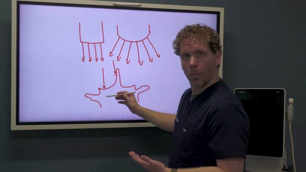
History Heuristics: Compression vs Stretch
- Joint/bone pain tends to worsen with compressive or provocative intra-articular motions (e.g., flexion, internal rotation). Patients with hip OA often hurt with axial loading or “grinding” positions.
- Soft-tissue pain (ligament/tendon) typically worsens with stretch (e.g., passive abduction aggravating adductor pathology).
- Nerve pain reproduces with tension tests (distribution-consistent radicular symptoms).
Range of Motion & Nerve Tension
- ROM: Flexion ≈120°; ER ≈40–60°; IR ≈30–40°. Early loss of internal rotation plus deep anterior/groin pain suggests intra-articular pathology.
- Nerve tests:
- SLR positive ~30°–70° for L5/S1 radicular pain; augment with ankle dorsiflexion (e.g., Bragard/Lasegue variants).
- Femoral stretch test (prone) for higher roots.
Intra-Articular Screens
- Scour test (quadrant): Axial load through the femur while sweeping arcs; anterior-superior quadrant is commonly symptomatic in labral disease. Sensitive but not perfectly specific—correlate with exam.
- FABER (Flexion–Abduction–External Rotation): Reproduces anterior hip or posterior buttock pain depending on pain source; add gentle overpressure with contralateral ASIS stabilization.
- Log roll: Passive internal/external rotation with the patient supine; highly specific in practice for intra-articular pathology when clearly positive.
Active Strength to Isolate Structures
Functional anatomy sharpens localization:
- Hip flexors:
- Knee extended (tests iliopsoas + rectus femoris).
- Knee flexed (biases iliopsoas, reduces rectus contribution).
Pain only with knee extended → suspect rectus femoris; pain with both → consider iliopsoas.
- Quadriceps vs rectus femoris:
- Straight-leg hip flexion activates all quads including rectus.
- Supported thigh with knee extension only emphasizes vasti over rectus.
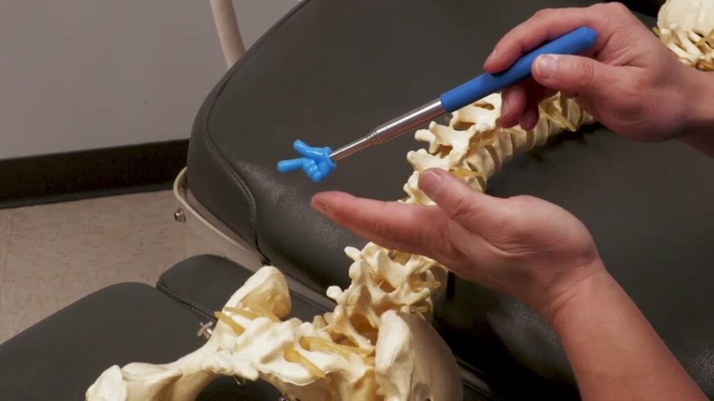
Surface Palpation: Landmarks That Matter
Palpation is highly sensitive—if you know what you’re pressing on.
- ASIS: Proximal sartorius/inguinal ligament; use the thenar eminence first to find bony prominences in higher BMI patients, then fine-tune with fingertips.
- AIIS: Proximal rectus femoris—often exquisitely tender; be gentle.
- Greater trochanter: Lateral pain is frequently gluteus medius/minimus tendinopathy; TFL/IT band lies more anterior and blends distally to Gerdy’s tubercle.
- Iliac crest (posterior-superior rim): Proximal gluteal tendon attachments can be tender.
- Ischial tuberosity (sits bone): Most tenderness is posterior-superior (proximal hamstrings, sacrotuberous ligament).
- History pearl: Hard surface sitting pain → hamstring/sacrotuberous bias. Soft surface sitting pain → think obturator internus (tension across the posterior ischium).
- Correlate palpation with diagnostic ultrasound to verify tissue injury and guide targeted injections/hydrodissection.
Clinical Takeaway
Think tissue first, then confirm with targeted maneuvers: compression for joints, stretch for soft tissues, tension for nerves. Combine ROM, scour/FABER/log roll, strength isolation, and precise palpation to localize the pain generator—and treat the right structure the first time.