Filter posts by category

Lower Extremity, Nerves

Differentiating Medial Knee Pain: Infrapatellar Saphenous vs. Inferior Medial Genicular Nerves

Differentiating Medial Knee Pain: Infrapatellar Saphenous vs. Inferior Medial Genicular Nerves

Medial knee pain is common in patients with osteoarthritis, ligamentous instability, and postoperative or overuse syndromes. Two frequent—but often conflated—pain generators live in the same neighborhood: the infrapatellar branch of the saphenous nerve (IPS) and the inferior medial genicular nerve (IMGN). Understanding how to find and treat each one can significantly improve outcomes.

Quick Anatomy Review

  • Saphenous nerve & IPS branch: The saphenous nerve originates from the femoral nerve and travels through Hunter’s (adductor) canal, providing cutaneous sensation along the medial knee, calf, and ankle. The infrapatellar branch is a small, recurrent sensory branch that innervates the anteromedial infrapatellar region—superficial, within subcutaneous fascial planes above the pes anserine tendons and superficial to the MCL.
  • Inferior medial genicular nerve (IMGN): A capsular branch accompanying the inferior medial genicular artery, curving around the medial tibial flare to innervate the inferomedial joint capsule. It sits deep to the MCL, adjacent to the tibial cortex.

Why They’re Easy to Confuse

Patients often report focal tenderness over the medial tibial plateau/infrapatellar area, where both IPS (superficial, cutaneous) and IMGN (deep, capsular) converge clinically. Palpation alone can be inconclusive; you may elicit tenderness over the pes anserine region, MCL, or along the saphenous track to the medial malleolus without confidently assigning the driver.

Ultrasound Roadmap

  1. Landmarks: Place the probe over the medial tibial plateau. Identify the tibial cortex as a bright hyperechoic line (the tibial flare). Superficial to cortex, you’ll visualize the MCL with linear fibrous architecture; superficial to the MCL are the pes anserine tendons.
  2. Find the IMGN (via its artery): Activate power Doppler and look for the inferior medial genicular artery at the tibial flare, just deep to the MCL. Adjust Doppler gain high enough to catch small-vessel flashes (too low and you’ll miss it; too high and you’ll get speckle). The nerve tracks with the artery—you may not always visualize the nerve, but the artery is your beacon.
  3. Locate the IPS branch: Scan superficial subcutaneous fascial planes over the anteromedial infrapatellar region, above pes anserine and the MCL. The IPS lies in these planes as small hypoechoic fascicles within the fascia.

Treatment Strategy: Layer by Layer

  • Superficial (IPS): For cutaneous, burning, or pinpoint medial infrapatellar tenderness, perform perineural hydrodissection of the IPS within the subcutaneous fascial planes. D5W (5–10 mL) is commonly used to separate fascial layers and down-regulate the irritated branch.
  • Deep (IMGN): For capsular, “inside the joint” ache with focal tenderness at the tibial flare, target the IMGN deep to the MCL, again using hydrodissection (≈5 mL D5W) around the artery-nerve bundle. This can reduce intra-articular–type pain and relieve entrapment at the capsular margin.
  • Adjuncts: Track tenderness along the saphenous route (Hunter’s canal to medial malleolus) to identify broader saphenous involvement. Combine with standard OA and instability care plans as indicated.

Clinical Takeaway

Think in layers: superficial cutaneous pain suggests IPS; deep capsular pain points to IMGN. Use ultrasound landmarks (tibial cortex → MCL → pes anserine) and power Doppler to confidently identify the IMGN via its artery, and treat each plane with targeted hydrodissection. Precise diagnosis plus minimally invasive perineural techniques can meaningfully improve medial knee outcomes.

Differentiating Medial Knee Pain: Infrapatellar Saphenous vs. Inferior Medial Genicular Nerves Read Post »

Lower Extremity, Nerves

How to Diagnose and Treat Low Back Pain: Differential Diagnosis

How to Diagnose and Treat Low Back Pain: Differential Diagnosis

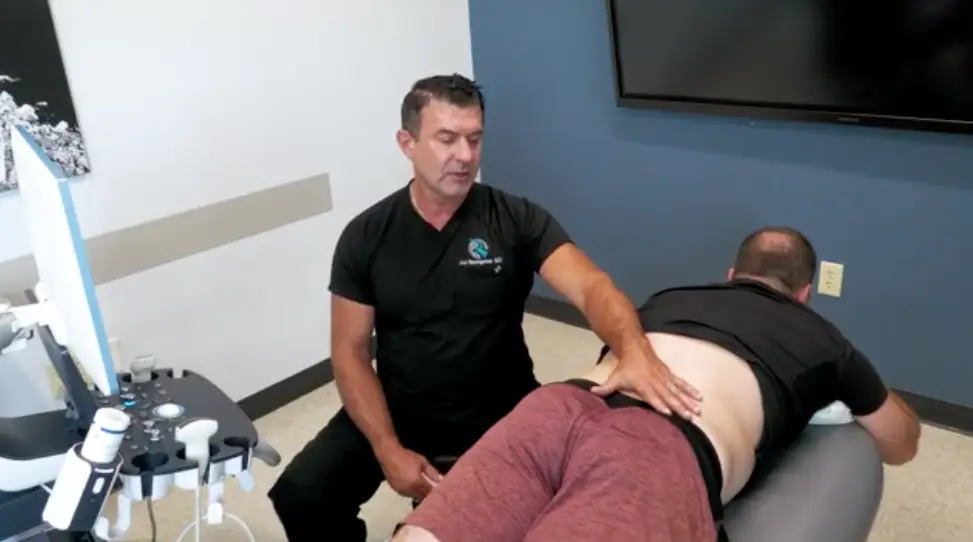
Low back pain is a common complaint among patients, and effectively diagnosing and treating it requires a nuanced understanding of its various causes and effective interventions. This guide provides a comprehensive overview of how to navigate the complexities of low back pain, from differential diagnosis to advanced diagnostic and treatment techniques.

How to Understand the Differential Diagnosis in Low Back Pain:
Understanding the differential diagnosis in low back pain is critical for effective treatment. It’s important to differentiate between actual spine pain and radicular spine pain, often stemming from conditions like degenerative disc disease and facet arthritis. Distinguishing between these can guide targeted interventions and improve patient outcomes.

How to Utilize Diagnostic Exam Techniques:
Diagnostic exam techniques are pivotal in identifying the specific cause of low back pain. By employing various methods, practitioners can discern between facetal versus dyspogenic pain sources, enabling the formulation of a more personalized and effective treatment plan. Mastery of these diagnostic skills is essential for healthcare providers.

How to Identify the Role of the Ileal Lumbar Ligament:
The ileal lumbar ligament plays a significant role as a pain generator in low back pain. Recognizing its role can guide targeted interventions, such as diagnostic injections, to alleviate discomfort. Understanding the anatomical structures involved in back pain is crucial for effective treatment.

How to Approach Thoracolumbar Fascia Pain:
The thoracolumbar fascia is a common site for pain due to its extensive connective tissue. Exploring the complex interplay between various anatomical structures and pain in this area can help in crafting more effective treatment plans. A deep understanding of the thoracolumbar fascia’s contribution to pain is essential.

How to Implement Diagnostic Injection Solutions:
Diagnostic injections are a valuable tool for identifying and treating sources of back pain. By strategically using injections, especially targeted at structures like the ileal lumbar ligament, practitioners can provide significant relief. This method serves as an important component of a comprehensive treatment plan.

How to Address Nerve-Related Pain:
Understanding how nerves contribute to pain is vital for diagnosing and treating back pain effectively. Focusing on nerve-related pain, including the role of nerves like the eli hypogastric, underscores the complexity of back pain and the need for a nuanced approach to treatment.

How to Enhance Diagnostic Accuracy with Ultrasound Skills:
Integrating ultrasound into the diagnostic process allows for a non-invasive examination of the back, providing real-time images of the spine and surrounding tissues. These advanced imaging techniques enhance diagnostic accuracy and guide treatment decisions, leading to better patient outcomes.

By focusing on these key areas, healthcare professionals can improve their management of low back pain, offering patients relief and a better quality of life. Understanding the intricacies of low back pain diagnosis and treatment is a journey, but with the right knowledge and tools, it’s one that can lead to significant benefits for both practitioners and patients alike.

How to Diagnose and Treat Low Back Pain: Differential Diagnosis Read Post »

Lower Extremity

Foot & Ankle Physical Exam: A Fast, Structured Walkthrough

Foot & Ankle Physical Exam: A Fast, Structured Walkthrough

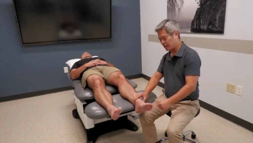
Dr. Wang lays out a practical, clinician-friendly foot and ankle exam using the classic flow: inspection → palpation → range of motion → special tests. Here’s the distilled playbook.

1) Inspection (standing and supine)

Standing (360° look):

  • Alignment & deformity: toe rotation, valgus/varus, hallux valgus, claw/hammer/mallet toes.
  • Arch/biomechanics: compare arches side to side. Use a quick “finger under the arch” screen; relative pes planus is often obvious visually.
  • “Too many toes” sign: from behind, seeing more lateral toes on one side suggests posterior tibialis dysfunction and medial arch collapse.
  • Heel rise test: during plantarflexion, a subtle lateral shift of the ankle/medial heel at end-range is normal; loss of this excursion suggests hindfoot/ subtalar instability.
  • Tendons/bursae: look for fusiform thickening of the Achilles and swelling in the retrocalcaneal bursa.

Supine:

  • Survey for edema (check pitting), erythema, ecchymosis (often migrates distally over days), and focal swelling over joints/tendons.

2) Palpation (think quadrants)

Patients often localize pain precisely—use that advantage.

Anterior:

  • Joint line (tibiotalar) vs extensor tendons (TA, EDL, EHL).
  • Tarsometatarsal (TMT/Lisfranc) region: anatomy is dense; identify the tender point, then correlate with imaging or ultrasound to map talus → navicular → cuneiforms → metatarsals.

Medial:

  • Deltoid ligament (proximal to malleolus) and sustentaculum tali (just inferior)—common tender spots.
  • Behind the medial malleolus (Tom, Dick, AN, Tom): TP (most commonly symptomatic), FDL, artery/veins/nerve, FHL. Trap TP against bone to provoke focal tenderness.
  • Spring ligament and navicular plantar-medial tenderness; plantar fascia origin just anteromedial to the calcaneal tuberosity.

Lateral:

  • ATFL (from lateral malleolus toward big toe)—hallmark tenderness after inversion sprain.
  • Sinus tarsi (anterior–inferior “divot”): deep ligament pain in repetitive inversion injuries (sinus tarsi syndrome).
  • Peroneals behind the malleolus; look for retinacular pain, popping/subluxation, and fibularis brevis insertion pain at the base of the 5th metatarsal (differentiate stress/“marcher’s” fracture vs Jones avulsion).

Posterior/Plantar:

  • Achilles: watershed zone 2–6 cm proximal to insertion is classic for tendinopathy; fusiform swelling is typical.
  • Plantar fascia (medial > lateral band), plantar plate tenderness (apply distal-to-proximal directed pressure), sesamoids (medial/lateral).

3) Range of Motion (ROM)

  • Dorsiflexion/Plantarflexion: posterior chain tightness vs anterior impingement; dancers may report posterior impingement in PF.
  • Inversion/Eversion: inversion stresses ATFL/CFL; eversion is less common but can be painful with deltoid injury.
  • Midfoot/forefoot torsion: assess pronation–supination mechanics.
  • Hallux MTP: screen for hallux rigidus/limitus (loss of extension most common).

4) Special Tests

  • Thompson test: prone calf squeeze → absent PF = Achilles rupture.
  • Anterior drawer: calcaneus forward on stabilized tibia → ATFL laxity.
  • Talar tilt (inversion stress): targets CFL.
  • External rotation stress test: stabilizes tibia, externally rotates foot → distal pain (and sometimes proximal fibular symptoms) suggests syndesmotic (“high ankle”) sprain.
  • Metatarsal torsion test: invert/evert forefoot while stabilizing midfoot—reproduces pain/laxity at TMT joints.
  • Intermetatarsal shear: isolate motion between adjacent metatarsals to detect intermetatarsal ligament sprain.
  • Metatarsal squeeze (Morton’s neuroma): ML compression of distal metatarsals with head stabilization → neuropathic pain/paresthesia.
  • Grind tests (axial load + circumduction): sensitive for MTP/IP arthropathy—start gently.

Pearls

  • Let patient-pointed tenderness guide you; the foot’s localization is often exact.
  • Map pain by quadrant, then confirm with ultrasound or plain films for joint/tendon differentiation.
  • Don’t miss posterior tibialis dysfunction, sinus tarsi syndrome, Achilles watershed tendinopathy, 5th metatarsal base fractures, and syndesmotic injury—they change management.

Foot & Ankle Physical Exam: A Fast, Structured Walkthrough Read Post »

Spine

Pelvic & Lumbar Nerve Entrapments: A Practical Guide for Persistent Low-Back and Pelvic Pain

Pelvic & Lumbar Nerve Entrapments: A Practical Guide for Persistent Low-Back and Pelvic Pain

Low-back pain is one of the most common complaints in clinical practice—and a surprising amount of it isn’t purely disc, facet, or SI-joint related. Peripheral nerve entrapments around the lumbar spine and pelvis often drive pain patterns that look “radicular,” resist standard care, and linger for years. In this quick guide—adapted from a live teaching session—we’ll tour the key posterior and anterior pelvic nerves, how they get trapped, and practical ways to find and treat them with palpation and ultrasound.

Posterior Pelvis: Meet the Cluneal Nerves

Why they matter. Superior, middle, and inferior cluneal nerves provide cutaneous innervation across the low back and buttock. They’re frequently irritated in the setting of lumbar/SI instability and facet degeneration—think “double crush”: one site at the spine/facet and another as the nerve crosses fascia or bone.

Superior cluneal nerve (SCN—especially the L3 branch).
The L3 SCN is the usual suspect. It’s commonly irritated:
• Proximally near the L5–S1 facet, and
• Distally as it passes a fibro-osseous tunnel over the iliac crest.
Patients may have focal tenderness along the crest and pain that tracks toward the greater trochanter—a helpful mental “target” because many posterior pelvic sensory branches visually and clinically “point” there.

Middle cluneal nerve (MCN).
This nerve traverses the posterior SI ligaments and the paraspinal musculature. The S1 branch takes a sharp turn beneath the PSIS, running over the posterior long SI ligament—a classic spot where tissue glide is poor and palpation is exquisitely tender. Hydrodissection here can instantly change a “disc-like” pain picture.

Inferior cluneal nerve (ICN).
A branch of the posterior femoral cutaneous nerve, the ICN emerges near the gluteal cleft and innervates the inferior-medial buttock. Its territory overlaps with pudendal branches, so patients with “sit bone” or rectal-adjacent pain often report aggravation when seated on hard surfaces.

Landmarking and technique cues.
Systematically mark the PSIS, iliac crest (carry the line forward; the whole crest matters), the lateral sacral border, and the ischial tuberosity. With those bony rails mapped, palpation-guided injections become straightforward; ultrasound refines the plan by visualizing facets, fascial tunnels, and the nerve as it dives or turns.

Anterior Pelvis: Iliohypogastric, Ilioinguinal, Genitofemoral & Friends

Iliohypogastric (IH) vs Ilioinguinal (II).
These travel between the internal oblique and transversus abdominis before getting more superficial. Two rules help:
• Trajectory around the crest:
  – IH tends to run 1–2 cm above the iliac crest.
  – II runs on the crest.
• Inguinal canal behavior:
  – IH stays above the canal (about 2 cm superior to the ASIS and the canal itself).
  – II enters and traverses the inguinal canal and supplies the pubic and proximal medial-thigh region.

Genitofemoral (GF).
Splits into a genital branch (often tracking with II through the canal) and a femoral branch that lies just superior to the femoral artery under the inguinal ligament (look ~1.5 cm lateral to the artery for tenderness).

Subcostal nerve.
Similar field to IH but typically 2–3 cm above it; has a lateral cutaneous branch between the mid-axillary line and ASIS.

Femoral & obturator nerves.
These are deeper and often best addressed with ultrasound:
• Femoral: identify the femoral artery, then look lateral for the nerve in the iliopsoas groove.
• Obturator: exits the obturator canal, then splits within the fascial planes between adductor longus, brevis, and magnus—a great target in chronic adductor strains and “sports hernia” patterns.

Lateral femoral cutaneous nerve (LFCN).
Classic meralgia paresthetica arises as LFCN crosses medial to the ASIS, under the inguinal ligament, and over the sartorius. Treat the triangle just distal/medial to ASIS, then track anteriorly to catch the bifurcation—posterior fibers run toward the fibular head, anterior fibers toward the VMO region.

The Knee’s Patellar Plexus: Don’t Forget the Rim

Anterior knee pain that worsens with kneeling isn’t always patellofemoral syndrome. The anterior femoral cutaneous branches, LFCN, and infrapatellar saphenous branches create a patellar plexus right along the patellar rim. Because these are superficial cutaneous nerves draped over bone, compression occurs at superior, mid-rim, and inferior points. Palpate the rim methodically; tender “snap-points” often respond dramatically to small-volume hydrodissection.

ACNES: Abdominal Cutaneous Nerve Entrapment Syndrome

When the GI workup is pristine but focal abdominal pain persists, think ACNES. Thoracic roots (T7–T12) travel between abdominal wall layers, then turn sharply through the linea semilunaris and rectus sheath to pierce the fascia via a small aponeurotic ring—a perfect choke point.
Clues: a fingertip-sized spot of maximal tenderness, a positive Carnett sign (pain remains or worsens when the patient tenses the abdomen), and immediate relief after a small diagnostic/therapeutic injection into the ring. Ultrasound helps you find the fascial exit; Doppler may show the companion artery.

TAP Blocks & Why Ultrasound Wins

A transversus abdominis plane (TAP) block spreads fluid between the internal oblique and transversus abdominis, bathing IH/II (and sometimes subcostal) along their course. You can approach more lateral (mid-axillary, over the iliac crest) or more anterior (near the ASIS), depending on where palpation and symptoms localize. Ultrasound confirms the three muscle layers and shows the hydrodissection plane in real time.

For the posterior pelvis, ultrasound is equally helpful:
• Visualizing the L5–S1 facet adjacent to the SCN
• Dissecting the posterior long SI ligament for MCN entrapment
• Tracking the pudendal course between the sacrospinous and sacrotuberous ligaments toward Alcock’s canal
• Identifying the sciatic and posterior femoral cutaneous nerves around the ischial tuberosity and quadratus femoris

Caudal Epidural with Dextrose: A Safe, Central “Reset”

As a complement to perineural work, a caudal epidural (performed under ultrasound by identifying the sacral cornua and entering the canal beneath the sacro-coccygeal ligament) can “centralize” pain and calm multiple irritated roots. Hyperosmolar solutions like dextrose have been studied for decades; many clinicians now use dextrose as an active therapeutic rather than just a carrier. Ultrasound and color flow confirm correct spread in the epidural space.

Take-Home Pearls

• If you can push it and reproduce the pain, it’s probably peripheral. Radiculopathy often isn’t tender to focal palpation; cluneal and cutaneous entrapments usually are.
• Map first, treat second. Mark PSIS, iliac crest, sacral border, ischial tuberosity, ASIS, and the inguinal ligament. Landmarks turn chaos into a protocol.
• Think in planes and tunnels. Fascia + bony edges + sharp turns = likely choke points.
• Use ultrasound to see the problem. It upgrades safety, accuracy, and patient confidence.
• Remember the “greater trochanter target.” Many posterior pelvic branches aim toward it—track pain patterns with that in mind.

Clinical content is for educational purposes for trained healthcare professionals. Patients should consult qualified clinicians before any procedure or treatment.

Pelvic & Lumbar Nerve Entrapments: A Practical Guide for Persistent Low-Back and Pelvic Pain Read Post »

Scroll to Top